Glossary
A
Acquired – Conditions caused by lifestyle factors, injury, or other conditions instead of being passed from a parent to their child (heritable condition).
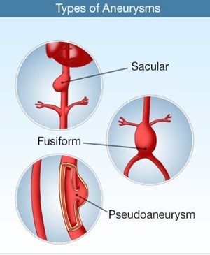
Aneurysm – A bulge or “ballooning” in the wall of an artery (Figure 1)
- A fusiform aneurysm is when the aorta is enlarged all the around the entire aorta. This is the most common type of aortic aneurysm.
- A saccular aneurysm is a focal outpouching of the aortic wall and is a true aneurysm involving all three layers of the aortic wall.
- A pseudoaneurysm (false aneurysm) is also called a contained rupture of the aorta and is due to either trauma, rupture of the aorta, or a penetrating aortic ulcer.
Angiography/Angiogram – An invasive procedure where intravenous contrast is injected into a blood vessel to see blood flow in a vessel using X-rays.
Aorta – The largest artery in the body which carries oxygenated blood from the heart to the body (Figure 2). The aorta is divided into anatomic segments: the aortic root; the ascending aorta; the aortic arch; the descending aorta; the abdominal aorta.
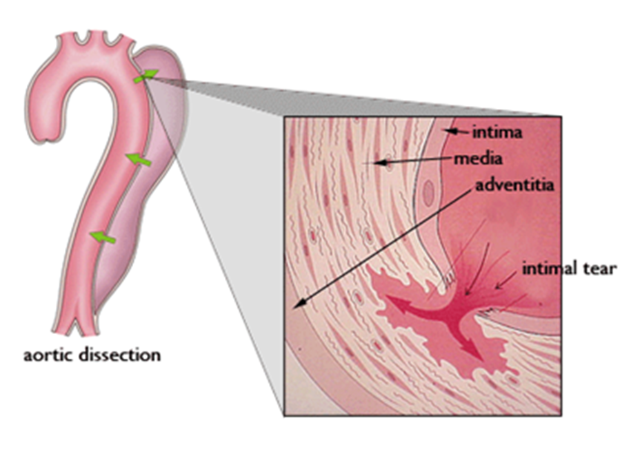
Aortic dissection – When a tear or split in the wall of the aorta occurs which allows blood to enter and flow in the aortic wall (Figure 3). This process can lead to aortic rupture or aortic bleeding, and it may interfere with the normal blood flow to organs or the extremities.
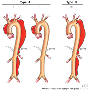
Aortic dissections are classified based upon the location of the tear or dissection in the aorta (Figure 4). In the Stanford classification, Type A dissections involve the ascending aorta and Type B dissections begin in the descending aorta. In the DeBakey classification, a type I dissection begins in the ascending aorta and extends to at least the aortic arch (and often throughout the entire descending aorta), a type II dissection is limited to the ascending aorta, and a type III dissection begins in the descending aorta.
Aortic Insufficiency/Aortic Regurgitation – A condition that occurs when the aortic valve does not close properly. This leakage (regurgitation) leads to blood flowing back into the heart after it has pumped blood to the body.
Aortic Stenosis – A thickening and narrowing of the aortic valve. This thickening prevents the valve from opening properly.
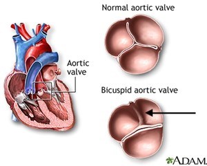
Aortic Valve – The heart valve that separates the main pumping chamber of the left ventricle and the aorta. Allows blood to flow normally from the heart’s left ventricle into the aorta when the heart pumps. It normally has three leaflets (Figure 5).
Arrhythmia – An irregular beating of the heart. The heart can beat too fast, too slow, or in an irregular pattern.
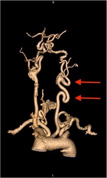
Arterial Tortuosity – A condition of the blood vessels characterized by elongation and severe twistedness (tortuosity) of the major arteries (often to the neck and brain) and may include the aorta (Figure 6).
Artery – A blood vessel that carries blood rich in oxygen from the heart throughout the body.
Atherosclerosis – A buildup of plaque (cholesterol) in the artery walls. This causes hardening of the arteries and may restrict blood flow to organs or to the rest of the body.
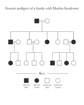
Autosomal Dominant Inheritance – A person has two copies of each chromosome, and two copies of each gene. If a condition is autosomal dominant, only one copy of the gene needs to have a mutation for the person to have the condition. For autosomal dominant conditions, there is a 50% chance the person’s children will also get the condition (Figure 7).
B
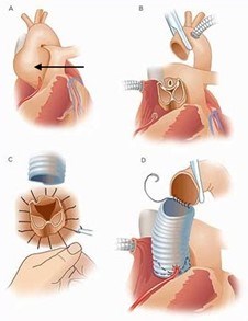
Bentall Procedure (or composite aortic root replacement) – Surgery that replaces the aortic valve, aortic root, and ascending aorta with an artificial valve and surgical graft (Figure 8).
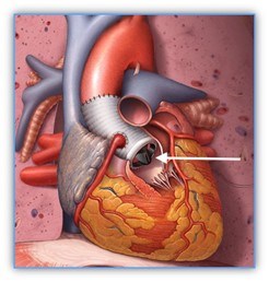
Bicuspid Aortic Valve – An abnormal aortic valve with two leaflets instead of three (refer to Figure 5, above).
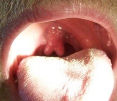
Bifid Uvula – A birth defect resulting in a split uvula, the fleshy extension hanging at the back of the palate above the throat (Figure 9).
Blebs – A blister-like enlargement in the lungs.
Blood Pressure – The pressure measured as blood is pumped through the arteries. Normal blood pressure is around 120 systolic (peak pressure in the arteries) over 80 diastolic (lowest pressure in the arteries).
Blood Thinner (anticoagulant) – A drug used to lessen the risk of formation of blood clots. Blood-thinners affect the clotting system in our bodies and prolong the time it takes for blood to clot.
Blood Vessels – A blood vessel is a tube that carries blood in the circulatory system. Blood vessels that take blood away from the heart are arteries. Blood vessels that take blood back to the heart are veins.
Bullae – Sacs, or abnormal enlargements, in the lungs. Bullae are larger than blebs.
C
Cardiac Catheterization – A procedure in which a catheter, a thin flexible tube, is passed into the right or left side of the heart. This procedure is used to diagnose heart conditions. It can also be used to treat heart conditions, like angina and heart attacks.
Cardiopulmonary Bypass – A procedure in which a machine takes over the function of the heart and lungs. It is used during surgery to maintain blood circulation and oxygenation to the body while surgeons operate on the heart and aorta.
Cervical-spine instability – An abnormality of the neck that may lead to dislocation of the spine and risk for spinal cord damage.
Chromosome – Genes contained in a specific area. Each cell has two of the same chromosomes; one from a person’s mother and one from their father.
Chronic Obstructive Pulmonary Disease (COPD) – A chronic lung disease that makes it hard to breathe. Less air than usual is able to flow in and out of the lungs.
Cleft Palate – An opening or split in the roof of the mouth that occurs when the tissue doesn’t fuse together during development in the womb.
Clubfoot – A condition in which a newborn’s foot or feet appear to be rotated internally at the ankle. The foot points down and inwards, and the soles of the feet face each other. It is known as talipes equinovarus (TEV) or congenital talipes equinovarus (CTEV).
Computed Tomography (CT) scan – A test that combines a series of x-rays to provide a detailed image of the body. When a dye is injected into the arteries, physicians have a detailed picture of the blood vessels.
Congenital – A disease, condition, or syndrome present at birth.
Connective Tissue Disorders – A group of disorders that affect the parts of the body that connect and support the structures of the body together. Several of these disorders are associated with an increased frequency of aortic aneurysms aortic dissections, such as Marfan syndrome, Loeys-Dietz syndrome, and Vascular Ehlers-Danlos syndrome.
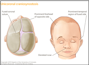
Craniosynostosis – A condition in which the bones in an infant’s skull grow together too early, causing problems with brain growth and head shape (Figure 10). This may be present in Loeys-Dietz syndrome.
C-section – In a Caesarean section, or C-section, surgery is used to deliver a baby.
D
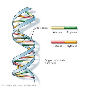
DNA – Long stretches of molecules that use a special alphabet to write instructions for the body (Figure 11).
Dissection – (Aortic Dissection) A tear in an artery wall. The inner lining rips open and blood enters the tear creating a false channel or false lumen (see Figure 3 above)
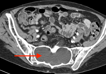
Dural Ectasia – Enlargement of the sac that surrounds the spinal cord, typically present in the lower lumbar spine and sacrum (Figure 12).
Dural Sac – The covering that surrounds the spinal cord, made of connective tissue.
Dyspnea – Shortness of breath.
E
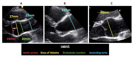
Echocardiogram – An imaging test that uses sound waves to see inside a person’s body (Figure 13). A transthoracic echocardiogram (TTE) is done by putting soundwaves over the chest wall. A transesophageal echocardiogram (TEE) is taken by inserting a tube into the throat and esophagus. No radiation is involved in this test. Also referred to as an ultrasound of the heart.
Electrocardiogram (EKG or ECG) – A non-invasive test in which the electrical signals generated by the heart are recorded. EKGs are often used to help diagnose heart problems, such as heart attacks or abnormal heart rhythms.
Endovascular Management – A less invasive procedure than open surgery in which the doctor accesses the aneurysm or dissection by a catheter, a thin flexible tube, inserted through a large artery in the arm or leg and pushed through the blood vessel to the aorta. TEVAR is the abbreviation for thoracic endovascular aortic repair.
F
First degree relatives – Parents, siblings, and children of the affected individual.
G
Genes – Contain instructions to make a protein or other molecule, which is used to build parts of the body. Genes are passed down from parents to their children.
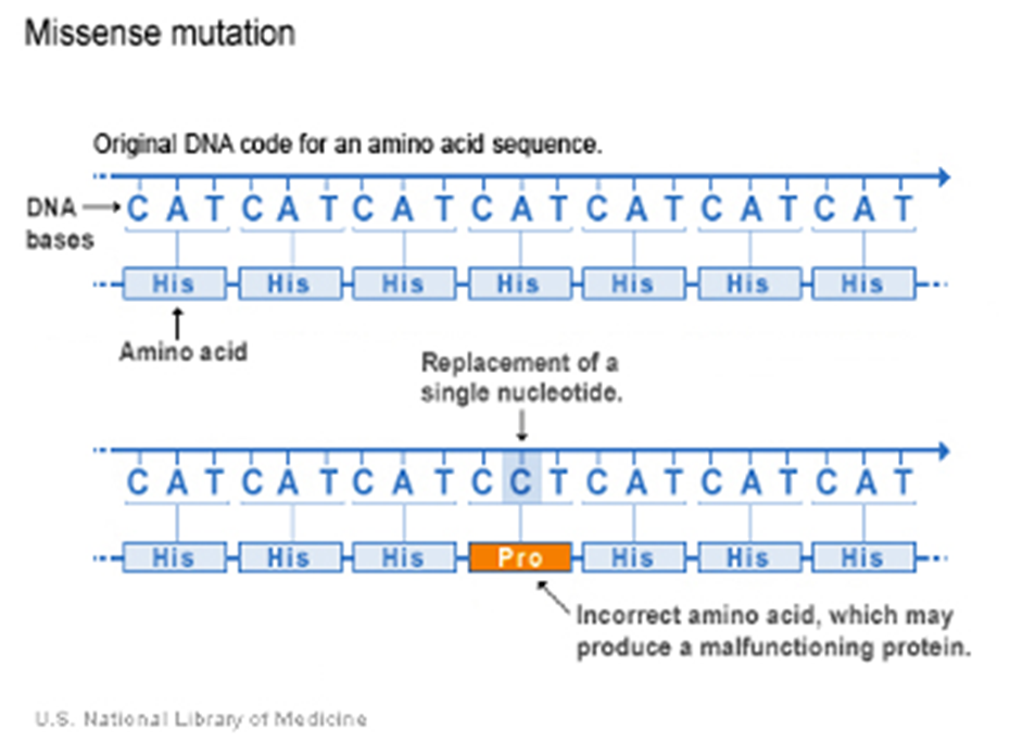
Gene mutation – Uncommon differences or changes in someone’s genetic code (Figure 14). These changes can disrupt the function of a gene, and may also affect how other genes work.
Genetics – Looks at how genes instruct the body to perform many different functions. Genetics also studies how genes can change, or mutate, and how genes are passed from parents to children.
Genetic testing – A medical test performed by collecting cells from the blood or saliva. This can be done to look for one single gene or many genes at one time.
Genotype – A person’s genetics, which go into determining their traits, or phenotype.
H
Heritable – Passed from parent to child.
Hernia – When an organ pushes through an opening in the muscle or tissue that holds it in place. For example, the intestines may push through a weakened area in the abdominal wall.
HTAD – This stands for Hereditary Thoracic Aortic Aneurysm Disease. HTAD is a group of conditions that cause problems with the aorta. The aorta is the major blood vessel that carries blood from the heart to the body. HTADs often run in families and can be passed from parents to children.
Hypertelorism – Abnormally increased distance between the eyes. Often part of Loeys-Dietz syndrome.
Hypertension – Also known as high blood pressure. Hypertension is typically diagnosed when blood pressure is above 130 systolic or 85 diastolic on two separate occasions.
I
Ischemia – An inadequate blood supply to a specific part of the body.
Isometric exercise – Exercise during which muscles contract against a fixed load, such as weight lifting, pushups, or pull-ups.
K
Kyphosis – Excessive outward front to back curvature of the spine, causing hunching of the back. The front to back curvature is as opposed to scoliosis, which is side to side curvature.
L
Loeys-Dietz Syndrome – A genetic connective tissue disorder due to a mutation in one of several genes. The severity may range from mild to life-threatening. Common signs and symptoms include widely spaced eyes, flexible joints, easily bruised skin, bifid (split) or broad uvula, tortuous arteries, and risk of aneurysm and aortic dissection.
Lumen – The opening inside of a vessel for blood to flow through. In aortic dissections, the aortic wall is split in two channels or lumens—the true lumen is the normal channel and the false lumen is the abnormal channel due to the split or tear in the aortic wall.
M
Magnetic Resonance Imaging (MRI) – A non-invasive test that uses magnetic fields and radio waves to provide a detailed picture of the body. MRA (Magnetic Resonance Angiogram) refers to using this technology to visualize blood vessels or the aorta.
Marfan Syndrome – A genetic connective tissue disorder. The severity may range from mild to life-threatening. Common signs and symptoms include a very tall and thin frame, flexible joints, eye lens dislocation, nearsightedness, collapsed lung, deformities of the chest wall, scoliosis, aortic aneurysm, and risk for aortic dissection.
Mitral Valve Prolapse (MVP) – A condition that occurs when the valve between the two left heart chambers (mitral valve) bows backwards into the left atrium when the left ventricle pumps. MVP is common in many syndromic aortic aneurysm disorders (such as Marfan and Loeys-Dietz). MVP may lead to leaking or regurgitation of the blood backwards into the left atrium when the heart contracts and, when severe, may require surgical repair.
Murmur – A sound made by blood moving in the heart and valves. Some murmurs may be harmless, while others may need further investigation with an echocardiogram to determine if a significant heart valve problem is present.
Myocardial Infarction (MI) – Commonly known as a heart attack. An MI happens when blood flow to one or more of the arteries supplying blood to the heart is blocked, causing damage to the heart muscle.
Myopia – Also known as nearsightedness. Near objects can be seen clearly, but far ones cannot.
P
Pathogenic – A gene mutation that is known to cause a disorder, such as Marfan syndrome, or a familial thoracic aortic aneurysm disease.
Pectus carinatum/excavatum – When the front of the chest is sunken (excavatum) or bulging out (carinatum) (Figure 15).

Phenotype – Observable or outward characteristics, or traits, that are based partially on a person’s genes. These can include things like eye color and height.
Pneumothorax – A collapsed lung often due to bursting of a bleb or bullae in the lung.
R
Retinal detachment – An emergency situation in which a thin layer of tissue at the back of the eye pulls away from the layer of blood vessels that provides it with oxygen and nutrients. Retinal detachment is often accompanied by flashes and floaters in a person’s vision.
Rupture – An aortic rupture is when the aorta bursts. This complication may occur with an aortic dissection or due to an aortic aneurysm.
S
Scoliosis – A sideways curvature of the spine that occurs most often during the growth spurt just before puberty.
Spontaneous mutation – A gene change that occurs naturally and is not passed from a parent to their child. If someone has a spontaneous mutation, they are the first in their family to have the condition.
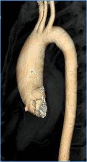
Stent Graft (TEVAR) – An endovascular procedure in which the doctor inserts a stent by way of an artery in the leg to treat an aortic dissection or aneurysm (Figure 16). TEVAR is the abbreviation for thoracic endovascular aortic repair.
Strabismus – Also known as cross-eyed. It results from weak control of one or more eye muscles.
Syncope – Fainting or loss of consciousness.
Syndrome – A group of symptoms that consistently occur together, or a condition characterized by a set of associated symptoms.
T
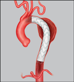
Thoracic aortic aneurysm (TAA) – When the aorta in the chest (thoracic aorta) is abnormally enlarged (Figure 17). Thoracic aortic aneurysms can be caused by genetic mutations, high blood pressure, inflammation (aortitis) hardening of the arteries (atherosclerosis), aortic dissection, or genetic (heritable) causes.
Turner Syndrome – A chromosomal abnormality in which a female is missing or partially missing an X chromosome. Turner syndrome is associated with an increased frequency of cardiovascular problems, including bicuspid aortic valve and ascending aortic aneurysm that increases the individual’s risk of aortic dissection.
V
Valve sparing procedure or valve sparing aortic root replacement – A surgery to replace an aortic root aneurysm during which the aneurysm is removed and an artificial graft is placed and the patient’s own aortic valve is spared and reimplanted within the graft (Figure 18).
Vascular Ehlers-Danlos Syndrome – A genetic connective tissue disorder due to a mutation in collagen. This condition leads to abnormalities of the skin, organs, and blood vessels. Common symptoms include varicose veins, abnormal bruising, sunken eyes, abnormal skin, collapsed lung, and blood vessel rupture and arterial dissection.
Vein – A blood vessel that carries blood from the tissues back to the heart.
X
X-Ray – A test that provides an image of the body.